
Many family doctors give a requisition or recommendation for a CT scan or an MRI without explaining the differences between those tests. As a result, patients, friends, and family members often ask us what the difference is between the two. How do they work and what is their purpose? Let’s review some basic points about these types of imaging studies.
A CT scan, also known as a CAT scan, is a rapid, painless examination that combines powerful X-rays and a computer to produce 360-degree, cross-sectional views of a body part or region. A CT scan can provide a lot of details about the body region that was examined.
CT scans are very good for bone injuries and certain soft tissues, such as the abdominal organs, lungs, and heart. It can also diagnose some diseases, such as cancer. Many times, intravenous contrast is used so that the radiologist can see the blood vessels very well.
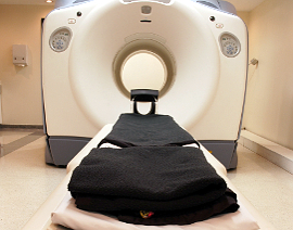
A CT machine looks like a big donut. To take the images, the part of the body being imaged passes through the machine. CT scans are widely used in ERs because this machine can take lots of images in less than 5 minutes. In fact, the actual scan time is usually less than 30 seconds.
Although this examination is painless, the main disadvantage of a CT scan is the radiation – too much repeated exposure can be harmful. That’s why it is not recommended for pregnant women or children unless necessary. The confined space can also create anxiety or claustrophobia for certain patients and very large patients sometimes can’t fit into the machine.
While a CT scan uses radiation, MRI combines a powerful magnetic field, radio waves, and an advanced computer system to produce accurate, detailed pictures of organs, soft tissues, bone (especially your bone marrow), and other internal body structures.
This type of imaging is best for soft tissue injuries, such as ligaments, tendons or muscles, spinal cord injuries, brain tumours, and other types of cancer since these anatomic structures contain magnetic properties. From a musculoskeletal point of view, MRI is the imaging of choice to detect and diagnose disc herniations, rotator cuff injuries, ACL tears in the knee, and even more conditions.
Because images are taken as a series of cross sections or “slices,” an MRI examination can take up 60 minutes, depending on which body region is being examined. The length of time can also depend on other factors, such as the type of machine used.
During the test, you must stay completely still; if you move, it will affect the quality of your images and it will be harder for the radiologist to read your images properly. Because an MRI examination is longer, patients usually get more anxious or claustrophobic than with a CT scan. Be aware that if you are going to have that test done, it makes very loud knocking sounds and it’s not pleasant at all!
Unfortunately, not everybody can have an MRI. The strong magnetic field can cause complications in patients with cardiac pacemakers, certain types of tattoos, and metal implants. Your doctor will perform a comprehensive patient screening procedure to determine if there might be any issues that make you ineligible for an MRI. If there are, you can still have a CT scan since it works with radiation instead of using a magnetic field.